Patient preparation is an important part of the success of capturing a 2D X-ray using a refurbished Planmeca ProMax machine from Renew Digital. You or a member of your dental staff should provide the patient with sufficient instruction and ensure that he or she understands the requests before moving forward with obtaining the image.
Basic Patient Instructions When Using Planmeca ProMax Equipment
Obtaining a clear and detailed image of your patient’s teeth, jaws, and gums requires cooperation from both the operator and the patient. The person obtaining the image should first instruct the patient to do the following before and during image capture.
- Remove all jewelry from the head and neck area, including piercings, if possible
- Close lips, refrain from swallowing, and remain still during exposure
- Place tongue on the palate to ensure that no air remains inside of mouth
- Stand up straight or follow the proper positioning protocol for wheelchair patients
Once the patient understands these steps, staff should start preparing the equipment.
How to Prepare Planmeca ProMax Equipment Before Taking Images
The first step requires a dentist or assistant to navigate to the equipment’s software and select the option for panoramic exposure. He or she should then follow these steps in order:
- Choose 2D dental, panoramic, and program type
- Choose the appropriate size for the patient
- If necessary, apply segmentation
- Press the arrow in the lower right corner to move the screen forward
The equipment is now ready for the patient positioning phase.
Steps for Proper Patient Positioning
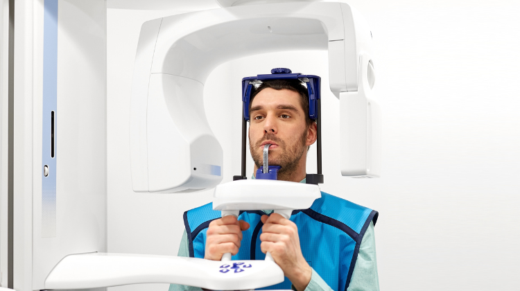
The staff person completing the imaging scan with Planmeca ProMax should start by adjusting the X-ray unit according to the patient’s height. The patient should then step towards the unit, place his or her chin on the chin rest, and grab the handles on each side. Next, the patient needs to bite into each groove on the bite plate. Staff should take over at this point and close the temple supports and set the patient’s head position. The Frankfort plane light will act as a guide and allow staff to get the proper position by moving the unit up and down.
Now the staff member should adjust the layer light between the patient’s canine and lateral incisor. After ensuring that the patient’s head lies straight against the midsagittal plane light, dental staff should select the correct size and shape for the patient’s jaw. As with the previous step, the staff person should click the go forward button in the lower right corner.
Obtaining Clear 2D Images
The first step in obtaining an image is to press the exposure button and hold it down. The image should appear immediately on an adjacent computer screen. With the procedure now complete, dental staff should guide the patient away from the Planmeca ProMax unit and back to the exam room. Keep in mind that it’s simple to set up the equipment to take specific images such as bitewing and panoramic.
Contact Renew Digital with Additional Questions
We understand there’s always a bit of a learning curve when working with new dental X-ray equipment. This is the reason we chose to include ongoing support in the purchase price of every piece of refurbished dental equipment that you buy from our company.