Although some patients may have concerns about radiation exposure during a dental appointment, the amount of radiation dentists use is so insignificant that many patients consider it an afterthought. When explaining dental X-ray machine radiation to patients, dental practitioners sometimes compare dental radiation dose to the amount of natural radiation a patient may receive from a few minutes lounging at the beach or traveling on an airplane. This helps patients better understand the risks of dental imaging vs. the rewards of maintaining proper dental health.
Advancements in Radiation Technology
Advancements in radiation technology have helped make dental X-ray machines much safer for patients and dental staff alike. For decades, dentists captured X-rays using traditional dental X-ray machines and film, then waited to develop the films before the images were available for diagnosis and treatment planning. Today, digital imaging technology has minimized the higher radiation doses often required with their film counterparts, as well as virtually eliminated the risk of retakes due to patient movement, poor quality results, or unwanted image artifacts.
Radiation Exposure in Dental vs. Medical Procedures
When compared to medical imaging, dental X-ray machines produce very little radiation, primarily because much more radiation is required to penetrate large areas of bone and tissue than is needed to penetrate dental anatomy. Additionally, the bones and skin of the mouth have low radio-sensitivity compared to larger parts of the body.For example, a 2D chest X-ray requires 50 μSv (micro Sieverts) of radiation, while a dental 2D panoramic X-ray requires only 10 μSv (micro Sieverts). Likewise, a medical CT scan of the maxilla and mandible can require up to 2,000 μSv (micro Sieverts) of radiation, as compared to only 36 μSv (micro Sieverts) for a similarly sized dental cone beam scan.
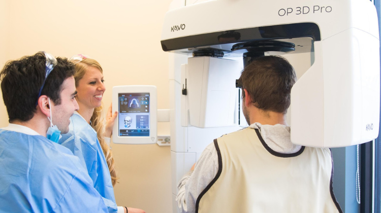
Three-Dimensional Scans in Dentistry
Thanks to the introduction of the cone beam dental X-ray machine, dentists only need very low doses of radiation to produce high-quality dental 3D images. Most modern dental cone beam systems can be collimated to capture only the region of interest, further reducing radiation dose.
Dental 3D scanners use cone beam technology and flat panel detectors, known as sensors, which are different than the higher-radiation based technology used in medical CT devices. Cone beam sensor technology has improved significantly over the years, requiring even less radiation to reconstruct dental cone beam images than the first generation models.
Dental Radiation and Cancer Risk
Radiation is often associated with cancer risk. If a patient is concerned about a 2D or 3D dental X-ray exam, they are likely worried about the risk of cancer. There is no scientific evidence that dental radiography has been shown to cause cancer. The fact that routine dental X-ray exams emit significantly lower radiation than even natural background exposures further supports that there is little to no risk associated with exposure from these types of exams.
Need additional tips for discussing dental X-ray machine radiation exposure with your patients? Renew Digital is happy to offer assistance at any time.