Since its recommendation by the American Academy of Oral and Maxillofacial Academy in 2012, 3D cone beam technology has become the standard of care for most dental professionals to obtain a complete picture when planning and placing dental implants. Cone beam computed tomography, or CBCT, provides you with a precise, relational view of the patient’s anatomy when planning implant procedures. 3D cone beam images, along with their associated 3D imaging software, allow practitioners to plan the treatment from implant through final restoration for the best and most predictable patient outcomes.
Benefits of Using 3D Cone Beam For Implant Planning
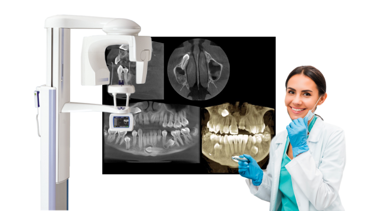
3D cone beam images allow you see patient anatomy in a three-dimensional view as opposed to only two dimensions. Traditionally, 2D periapical and panoramic radiographic images have been used to visualize and plan dental prostheses. The main disadvantage of these 2D images, however is that the buccolingual width of the alveolar bone and the location of the inferior alveolar nerves cannot be accurately assessed. This lack of information can often lead to implant positioning errors.
The use of 3D cone beam images during implant planning uses multiplanar and 3-dimensional volumes to determine the exact height, width and ridge anatomy of the alveolar bone and precise location of the alveolar nerve. It also helps define the relationship of edentulous sites with adjacent anatomy to ensure the greatest stability. Based on information obtained from the 3D cone beam images, the practitioner can determine if pre-prosthetic surgery such as bone grafting or sinus lift is needed prior to implant placement.
Benefits of 3D Imaging Software For Implant Planning
The 3D cone beam image itself is further enhanced by the powerful 3D imaging software to manipulate and manage the scan. Leading 3D imaging software included each 3D cone beam system provides practitioners with tools such as the ability to make annotations, adjust the window and level, and zoom in on specific areas.
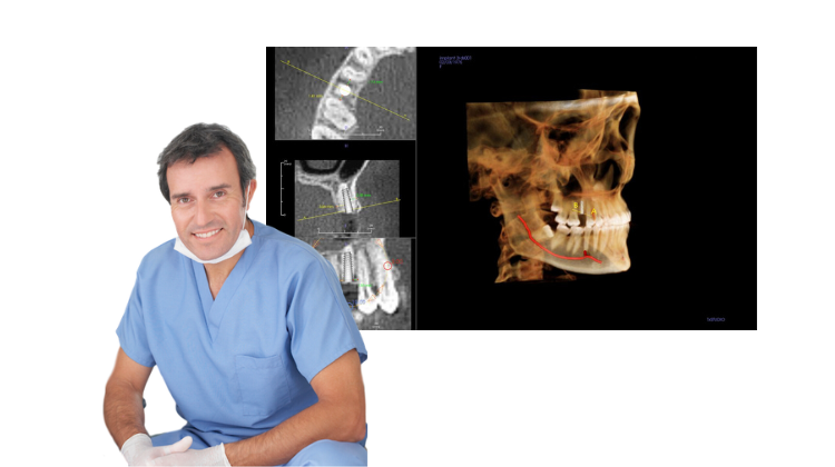
3D cone beam volumes can be exported into compatible implant planning software where you can take measurements, trace nerves, and evaluate bone density and regional anatomy. You can then select the shape, length, size, of the implant – or choose the exact implant from a comprehensive library of implant manufacturers – for even greater precision. With additional third-party software, you can even create surgical guides and virtual 3D models to further streamline implant planning.
Prosthetic-based implant planning software will utilize the full digital workflow, planning the treatment from surgery through restoration to ensure proper fit and function. Images can be shared with patients and restorative practitioners for comprehensive treatment planning and enhanced patient and referral communication.
Post-Operative Imaging Protocols
When following up on implant procedures, practitioners typically follow the ALARA (As Low as Reasonably Achievable) principle for radiation dose, and utilize 2D images, such as periapical, bitewing or panoramic images for post-operative care. However, If the implant is symptomatic or bone loss is present in adjacent anatomy, additional 3D cone beam scans may be required. Having a dual-purpose panoramic/cone beam dental x-ray machine in your practice for this purpose is a good plan for managing post-surgical follow-ups. That way, practitioners can choose the modality that best fits each patient’s unique post-operative imaging needs, while minimizing their radiation exposure.
Are You Ready for an Upgrade?
If you’re a dentist, dental specialist or oral and maxillofacial surgeon that would like to be more accurate and efficient during dental implant placement, we urge you to consider adding 3D cone beam equipment to your practice. Buying refurbished dental equipment helps to make it more affordable for you while also increasing the quality of service you provide to patients. Contact Renew Digital today for more information.