There are many benefits to using extraoral bitewings in your dental or dental specialty practice. Less cross-contamination by minimizing intraoral X-ray methods makes for a safer workplace, especially when considering the current global pandemic. Plus, you can often get the same high-quality images as intraoral X-rays for accurate diagnoses. You and your patients will feel peace of mind from the low radiation dose, plus you’ll all benefit from more efficient patient care.
If you’re ready to add extraoral bitewings to your practice, but unsure how to start, check out the guide below.

Types of Extraoral Bitewings
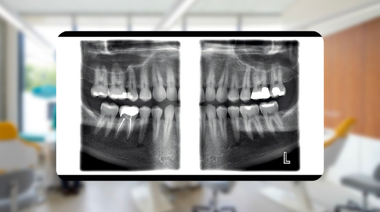
Dental X-ray machine manufacturers have developed two general types of extraoral bitewings: segmented and diagnostic. Some models capture only segmented extraoral bitewings. These images are created by taking a regular digital panoramic X-ray and segmenting out two bitewings. This bitewing view captures just beyond the apexes to beyond the opposing apexes. Diagnostic extraoral bitewings, offered by leading manufacturers such as Planmeca, however, incorporate a complex movement during panoramic rotation, following the curvature of the mandible during image capture. Bitewings are then separated out on a panoramic image-like view, but are often far more accurate than segmented extraoral bitewings.
How Extraoral Bitewings Work
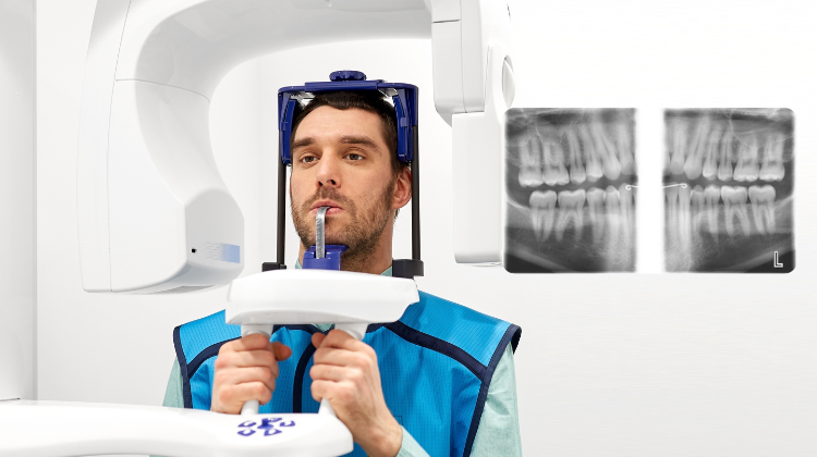
Some panoramic dental X-ray machine manufacturers offer an extraoral bitewing setting as part of the standard or optional panoramic X-ray programs. In these bitewing-enabled dental X-ray machines, the beam of the X-ray is placed perpendicularly, aligning with the long axis of your patient’s teeth. Image programming settings identify patient size and dental arch shape so the tube head moves closer to the patient, reducing the distance between the radiation source and the image. The extraoral bitewings are then automatically captured from canines to third molars and from crowns to apices.

Comparison to Intraoral Bitewings
Often, after reviewing traditional intraoral X-rays, many dentists tell patients “The good news is, we didn’t see anything! The bad news is, we didn’t see anything!” The patient is then sent home with a “Watch.”
In many cases, diagnostic extraoral bitewings provide more anatomical detail than their intraoral counterparts. The 360 degrees of rotation applying exposure through the mandible in panoramic imaging vast improves the paralleled image to the dentition which is captured by film, phosphor plates, and/or sensors. In conclusion, intraoral images can be sharper (often 20 Lp/mm) in contrast, but extraoral images (9.6-11.4 Lp/mm) have more depth of field.
Using Extraoral Bitewings to Detect Caries
In the past, dentists have used intraoral bitewings for caries diagnoses. Today, panoramic dental X-ray imaging technology has improved to a level where extraoral bitewings may provide superior diagnostic capabilities for diagnosing interproximal caries and bone loss than intraoral bitewings. Studies have shown that recent technology enhancements have improved extraoral bitewing image quality but dentists should still use the ALARA principle when prescribing dental radiographs.

Add Extraoral Bitewings to Your Practice!
If you're considering adding extraoral bitewings to your practice, Renew Digital can save you 30-50% off new list prices of new dental panoramic X-ray machines. Take a look at our wide range of panoramic dental X-ray machines enabled for extraoral bitewings today. Then, call us at 888-246-5611 or send a message to one our Sales Representatives. We can help you narrow down your choices to find the best unit for you, and offer current availability and pricing information.