If you're an endodontist, you already know how cone beam dental technology can benefit your practice. When you use cone beam computed tomography (CBCT), you can diagnose patients more effectively, plan treatments more accurately, and improve outcomes. Although cone beam dental technology has become the standard of care for most modern endodontists, many practitioners still have questions about the best way to use CBCT in their practices. We’ve put together some of the most frequently asked questions below.
When Should Cone Beam Computed Tomography be Used?
In 2015, the American Association of Endodontics (AAE) and the American Academy of Oral and Maxillofacial Radiology (AAOMR) issued a joint statement outlining the use of cone beam dental technology in endodontics. The statement defined specific recommendations for when cone beam should be considered, highlighting that not all endodontic treatment or diagnoses require cone beam dental images.
ALARA, the acronym for As Low as Reasonably Achievable, is the fundamental principle for diagnostic radiography. According to the AAE and AAOMR, clinicians should only use cone beam CBCT imaging when the diagnostic requirements cannot be adequately met by lower dose conventional dental radiography. Further, they stressed that CBCT must not be used for routine endodontic diagnosis or for screening purpose in the absence of clinical symptoms. Patient history combined with a comprehensive clinical examination must first justify the use of CBCT by demonstrating that the benefits outweigh the potential risks.
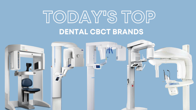
What Type of Cone Beam Dental Equipment Should my Endodontic Practice Consider?
For most endodontic applications, limited or focused field of view CBCT is preferred over large volume cone beam dental imaging. In general, the smaller the scan volume, the higher the resolution of the image and the lower the effective radiation dose to the patient. Increased resolution helps improve the diagnostic accuracy of endodontic-specific visualization of small features such as canals, lesions, and other anatomy.
What are the Advantages of Cone Beam Dental Imaging in Endodontics?
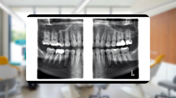
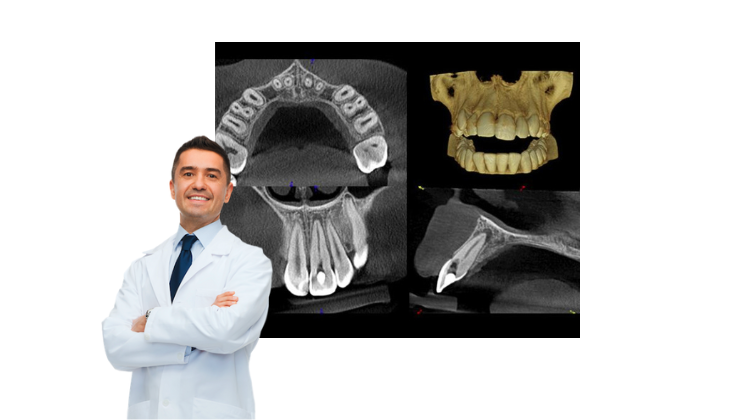
The most significant advantage of CBCT in endodontics is that it displays anatomic features in three dimensions compared to only two dimensions with intraoral and panoramic images. Cone beam systems reconstruct the imaging data in true spatial relationships. This multiplanar reformation allows practitioners to segment the data into cross-sections to highlight specific anatomic regions for faster and more accurate diagnoses.
In addition, most 3D imaging software provides enhancement tools such as zoom magnification, window/level adjustments, annotations, and measurements to further streamline diagnoses. Combining these tools with high-resolution cone beam dental images, CBCT technology provides clinicians with an unparalleled visualization of complex relationships and boundaries between teeth and their associated pathology and anatomic features within the alveolus and jaws, such as the maxillary sinus and mandibular canal and foramen.
How Do You Learn More About Using 3D Cone Beam Dental Technology?
Cone beam dental technology can help your practice to attract new patients, and it can improve the care you offer to existing patients. Cone beam equipment has become increasingly more affordable in recent years, and even more so when considering pre-owned alternatives. To learn more or to find CBCT equipment for your practice, look through our site or contact us directly. At Renew Digital, we focus on quality pre-owned imaging equipment that can help make your practice more successful while saving you money.