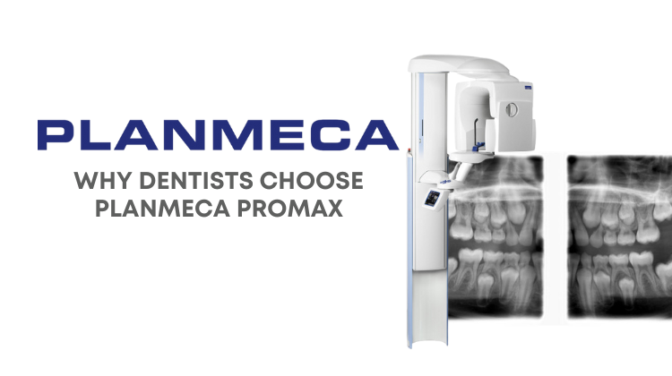
Cone beam dental scans have become the gold standard of dental imaging throughout the past two decades. They are especially helpful in the oral and maxillofacial surgery practice because they allow you to more effectively visualize dentition and dental anatomy without distortion and in relationship to other craniofacial structures. This helps you to more effectively and efficiently diagnose conditions, plan treatment, and communicate your treatment recommendations to your patients. All of these benefits ultimately lead to better surgical outcomes and higher patient satisfaction.
There are myriad different uses and applications for dental cone beam scans in the OMS practice. Let’s take a look at just some of them below.
Implants
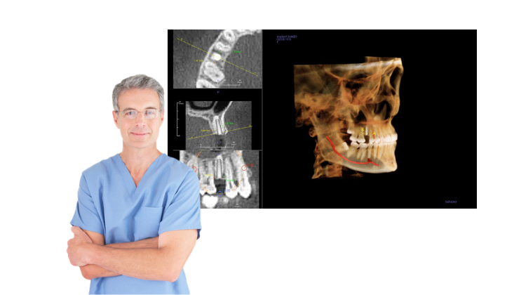
One of the most common and practical uses for a 3D cone beam imaging in an OMS practice is for placing dental implants. Dental cone beam images can help you more effectively plan implant treatment from start to finish. A dental cone beam scan can help determine the quality and quantity of the patient’s bone to see if implant placement is feasible and possible, help you accurately trace alveolar nerves and determine proximity to other teeth and dental structures in a non-invasive manner. Implant libraries and other implant planning tools can help surgeons better visualize implant placement along with final restorations, often resulting in greater treatment acceptance. Follow up dental cone beam scans can also help monitor implants post-operatively for more successful treatment outcomes.
Surgical Guides
Dental cone beam technology allows OMS practices to quickly and easily create computer generated surgical guides for placing multiple dental implants within a single arch. This enables surgeons to provide prosthetically-driven implant placements, enhancing surgical accuracy and efficiency while minimizing post-operative patient discomfort.
Impactions
A CBCT scan can show you the exact position of impacted teeth within the alveolar bone and in relation to other teeth, allowing you to thoroughly visualize and plan treatment before you begin surgery. This reduces staff surgical time, while improving patient outcomes with less anesthesia and post-operative pain and swelling. Dental cone beam images help you better determine the juxtaposition of third molar roots and more precisely communicate treatment concerns with patients and their families.
TMJ
With cone beam dental scans, you can assess morphological and arthritic changes in the temporomandibular joint and surrounding bony structures. In addition, you can more easily view and diagnose TMJ fractures, erosions, infections, and other anomalies previously undetectable on traditional 2D panoramic images due to overlapping anatomical structures.
Sleep Apnea
Measuring oral nasal airways and determining 3D volumes is made easier with cone beam dental technology. Fabricate appliances and use subsequent CBCT scans to evaluate if airway measurements or volumes have changed.
Pathology and Trauma
One major benefit of CBCT scans is the ability to pre-plan your surgery. You will be more confident with your approach, have a quicker surgery, and there will be less surgical swelling and discomfort for the patient.
Before the patient enters the surgical suite, you look at lesions such as large jaw cysts, see if buccal and lingual cortices are intact, evaluate teeth or nerve canal displacement, and get exact measurements of the bony volumes.
For dental trauma such as tooth avulsions, luxations, and fractures, a 3D cone beam can aid your diagnosis better than a 2D radiograph. It helps you to visualize alveolar fractures so you no longer have to feel along the ridge for movement.
Surgical Endodontics
Avoid engaging in exploratory surgery by using cone beam dental scans preoperatively. You can review the volume and see where the pathology has originated, as well as determine if it extends into the sinus. You can assess whether or not you should retreat canals, perform apicoectomy, or remove the tooth. Dental cone beam scans help provide the basis for a more definitive treatment plan for optimal patient care.
Diagnoses and Treatment Planning
When diagnosing and treating surgical cases, traditional 2D radiographs can be misleading. Recurring eruption cysts, or the fusion of impacted maxillary second and third molars might not show up on a 2D image. By getting another view with a 3D cone beam scan, you can ensure you are seeing exactly what is going on so you can make the best diagnosis and avoid unnecessary surgeries.
Bring 3D Cone Beam Technology to Your OMS Practice
Shopping with Renew Digital allows you to bring the advanced technology of 3D cone beam imaging to your oral and maxillofacial surgery practice at 30-50% off the cost of buying new dental cone beam equipment. Plus, your purchase includes installation, training, and a comprehensive full replacement part and onsite service warranty. Call us today at 888-246-5611 or send us a message online to learn more about our available units and pricing.