Digital ceph X-rays have come a long way since their inception a few decades ago. Modern digital technology makes taking and managing cephalometric X-rays faster and safer than ever before. Take a look at just a few important technology improvements in digital cephalometric X-ray design and function to help make practices more efficient and improve patient care.
Software Tools and Features
Today’s digital imaging software provides tools that allow for 1:1 measurement of ceph X-rays, which is a concern for many dental professionals migrating from analog systems. In addition, modern imaging software allows you to create serial superimpositions with cephalometric tracing. The ability to compare and contrast those images can be one of the most effective ways to measure growth, and these images can also help you see how the mouth is responding to treatments such as orthodontics. Plus, advanced treatment simulation modules available in some software packages, allow you to diagnose, plan, and present cases using 2D digital ceph X-rays, improving case acceptance.
“One-Shot” Cephalometrics
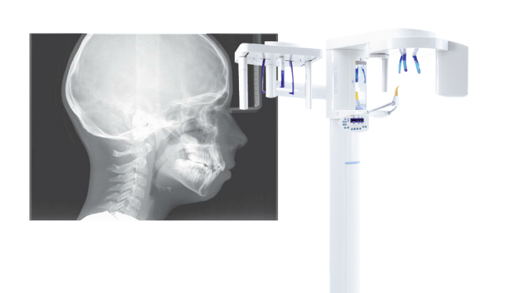
At first, digital cephalometric images were captured by scanning the patient twice, and then “stitching” two narrower images together to make a large cephalometric image. This dual capture method could result in seams between the two images as well as image distortion due to patient movement. Over the years, several dental X-ray manufacturers have introduced “One Shot” cephalometric models. Much like analog X-ray units, “One Shot” cephalometrics captures the entire area in a single pulse of radiation, minimizing risk of exposure, streamlining capture time, eliminating imaging seams and reducing motion artifacts.
Left or Right Configuration
Many practices don’t realize that flexible cephalometric X-ray configuration is important. Let’s face it, ceph X-rays are large and sometimes you have to be creative in your room design to get a cephalometric X-ray to fit. To help minimize this frustration, manufacturers feature models that allow for either a right or a left configuration. That means that the ceph X-ray attachment could be affixed to the right or the left of the panoramic X-ray machine based on spatial needs. If space is a consideration for your practice, make sure you ask about ceph orientation options.
3D and Cephalometrics
The introduction of large field of view cone beam systems have provided yet another way for practices to capture cephalometric images. You simply capture a large field of view 3D scan, then extract a 2D cephalometric image from the volumetric CBCT data. That way you get all of the records and patient information that you need in one convenient scan – a great solution for orthodontic or surgical practices. In addition, some X-ray systems today have the added benefit of a dedicated 2D panoramic/ cephalometric X-ray machine combined with a 3D sensor, giving you the flexibility of capturing 3D scans or traditional 2D images, based on your patient and practice needs.
Renew Digital offers a large selection of certified pre-owned 2D and 3D cephalometric options backed by a comprehensive parts, labor and support warranty. Plus, our knowledgeable sales representatives can help you find the perfect ceph X-ray for your practice. Take a look at our wide range of cephalometric systems online. Contact us today to learn more!